How is coarctation of the aorta treated?
How is coarctation of the aorta treated?
Twenty years ago, surgery was the only treatment available for aortic coarctation. Surgery is still considered the gold standard, but today treatment options for adults with this condition also include balloon angioplasty, stenting, stent grafting, or hybrid repair (a combination of open surgery and stent grafts). The choice of treatment is based on the individual's overall health, the size and severity of the coarctation, associated aneurysm or valve disease, and its precise location.
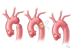
End-to-end anastamosis
An individual who is diagnosed with coarctation of the aorta should be under the care of an experienced congenital heart specialist at a major medical center. A cardiac surgeon experienced in the procedures used to treat this condition is best able to determine the optimal timing and choice of treatment.
Surgery
When the coarctation is relatively small, the surgeon can remove the narrowed section of aorta and re-join the two ends. This is called an end-to-end anastamosis and is often the best surgical option to treat the condition.
Other surgical options include various types of bypass surgery in which a graft is stitched onto the aorta to divert blood around the area of the defect. In patients with valve disease, a bypass can be performed in combination with a valve repair or replacement procedure.
Coarctation of the Aorta and Endocarditis
If you have had heart surgery or angioplasty to repair a defect, your doctor will prescribe preventive antibiotics for you to take before certain medical procedures for at least 6 months after the repair procedure to reduce the risk of infective endocarditis. Your doctor can provide specific guidelines about when to take antibiotics. According to the American Heart Association, there are insufficient data to make recommendations for continuing preventive antibiotic therapy longer than 6 months.
Angioplasty with or without stenting
Angioplasty is another option for treating coarctation of the aorta, and at some medical centers, it is the preferred treatment. The procedure is similar to angioplasty for coronary artery disease: a long, thin tube (a catheter) with a balloon on the end of it is passed into the aorta through the blood vessels, entering at the groin. When the catheter reaches the coarctation, the balloon is inflated to expand the aorta. When the narrowed area is fully expanded, the balloon and the catheter are withdrawn.
A small metal mesh tube called a stent may be placed at the site of the coarctation to keep the aorta open after the balloon is removed. The current trend is to recommend stenting in cases of severe, long areas of coarctation and in cases when differences in blood pressure persist after angioplasty alone. Stenting appears to lower the risk for aneurysm formation and rupture of the aorta compared with balloon angioplasty alone.
When the coarctation has an associated aneurysm, the repair should be performed with a stent graft (a cloth covered stent).
Recurring Coarctation
Following treatment for coarctation of the aorta, the late complications include recurrence of the coarctation (narrowed again), aneurysm (ballooning of the vessel), or pseudoaneurysm (a contained leak within the aorta wall).After surgical treatment, the risk of recurrence is between 5 and 10 percent. After angioplasty or stenting, the risk of recurrence is between 11 and 15 percent. However, long-term data on outcomes, risks and complications associated with angioplasty and stenting are not yet available.In the event that any late complications occur, evaluation and treatment at an experienced Aorta Center - that provides all types of treatments from stenting to redo surgery - is critical to achieving the best outcomes.
Follow-up Care
People who have undergone treatment for coarctation of the aorta should be under the ongoing care of a congenital heart disease specialist. Regular visits that include measurements of blood pressure and a clinical exam will be required every year. In addition, Doppler ultrasound and MRI tests should be performed at regular intervals, usually every two to five years.People who have normal blood pressure after treatment likely will have no activity limitations. Individuals who continue to have high blood pressure may be advised to avoid certain strenuous activities or sports.
